Figure 4.
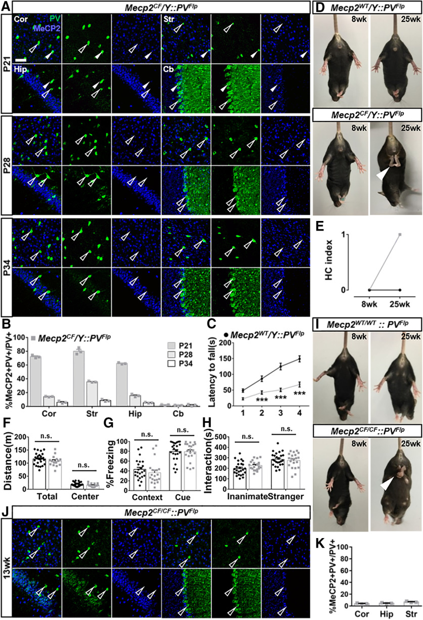
PV-specific Mecp2 inactivation mediated by PVFlp. A, B, Gradual loss of MeCP2 immunostaining in PV+ cells in cortex (Cor), striatum (Str), hippocampus (Hip), and cerebellum (Cb) in Mecp2CF/Y::PVFlp. A, Representative images. Scale bar, 50 μm. B, Quantification. n = 3. C–H, Behavioral performance of Mecp2WT/Y::PVFlp and Mecp2CF/Y::PVFlp littermates in rotarod test (C), HC (D,E), open field test (F), fear conditioning (G), and 3-chamber social test (H). Mecp2WT/Y::PVFlp, n = 26, 26, 24, 23, and 25; Mecp2CF/Y::PVFlp, n = 21, 21, 17, 20, and 21. D, I, Representative photographs HC phenotype in male (D) and female (I) when Mecp2 is inactivated in PV+ cells. White arrowhead indicates clasping hindlimb. J, K, Loss of MeCP2 expression in PV+ cells in the Cor, Str, Hip, and Cb of Mecp2CF/CF::PVFlp females. J, Representative images. Open arrowheads indicate PV+ cells without MeCP2. Filled arrowheads indicate PV+ cells with MeCP2. K, Quantification. n = 3. Data are mean ± SEM. Dots and squares represent data from individual mice. n.s., Not significant at p > 0.05, ***p < 0.001. For more statistical details, see Table 2.

