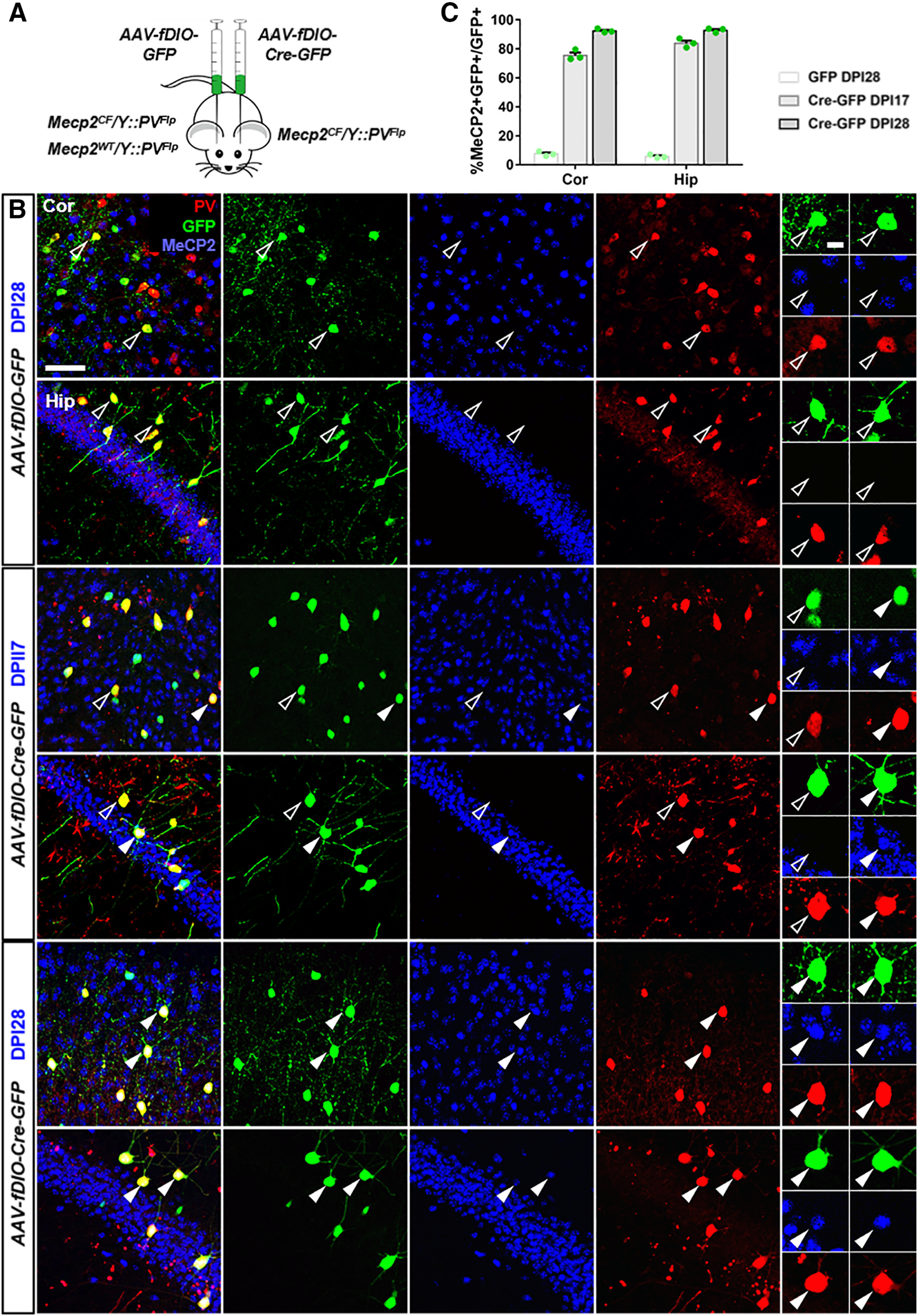
Figure 6.

Virus-mediated Mecp2 restoration in PV+ neurons. A, Experiment scheme. AAV-fDIO-GFP was injected into primary visual cortex or hippocampus of Mecp2CF/Y::PVFlp on one hemisphere, and AAV-fDIO-GFP-Cre on the other hemisphere. As a control, AAV-fDIO-GFP was injected Mecp2WT/Y::PVFlp to label WT PV+ neurons. B, C, Immunostaining confirmation and quantification of gradual Mecp2 restoration in AAV-fDIO-Cre-GFP-infected PV+ neurons. B, Top, AAV-fDIO-GFP-infected PV+ neurons in Mecp2CF/Y::PVFlp. Middle, Bottom, AAV-fDIO-Cre-GFP-infected PV+ neurons at 17 or 28 d post injection (DPI) in Mecp2CF/Y::PVFlp. Open arrowheads indicate GFP+ cells without MeCP2. Filled arrowheads indicate GFP+ cells with MeCP2. Panels on the right, High magnification views of representative cells. Scale bars: low magnification, 50 μm; high magnification, 10 μm. Data are mean ± SEM. Dots represent data from individual mice.
