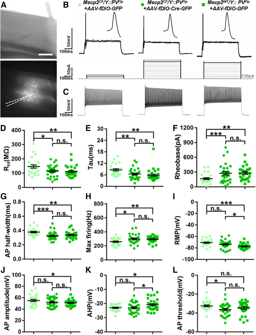
Figure 7.
Functional rescue by Mecp2 restoration in PV+ neurons in the primary visual cortex. A, Representative recording site imaged under infrared and fluorescent modes. Dashed lines in fluorescent image indicate recording pipette. Scale bar, 500 μm. B, Representative recording traces show APs evoked at rheobases; 10 pA depolarizing current steps were injected, and only the responses at rheobase and one step before it were shown. C, Representative recording traces showing APs at maximum firing rate. D–H, Rescued input resistance (Rin), membrane time constant (tau), AP half-width, rheobase, and maximum firing rate. Maximum firing was recorded as the firing rate at plateau. I–L, Increased resting membrane potential (RMP) and AP amplitude were partially rescued by Mecp2 restoration. Increased afterhyperpolarization potential (AHP) was not rescued. AP threshold was unaffected by Mecp2 inactivation. Data are mean ± SEM. Dots and squares represent data from individual recorded cells. n.s., Not significant at p > 0.05, *p < 0.05, **p < 0.01, ***p < 0.001. For more statistical details, see Table 1.

