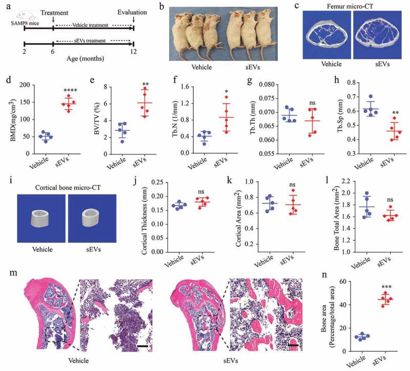
Figure 2.

hESC-sEVs treatment prevents age-related bone loss in SAMP8 mice.
(a) Experimental design for testing the effect of hESC-sEVs on preventing age-related bone loss: 6-month-old male SAMP8 mice were randomized to either vehicle or hESC-sEVs treatment for 6 months. (b) Representative images of aged male mice applied with vehicle or hESC-sEVs. The mouse images were taken after 6 months of treatment. (c) Representative micro-CT images of bone microarchitecture at the femur. (d–h) Quantification of micro-CT derived bone mineral density (BMD; mg/cm3) (d), bone volume fraction (BV/TV; %) (e), trabecular number (Tb. N; 1/mm) (f), trabecular thickness (Tb. Th; mm) (g), trabecular separation (Tb. Sp; mm) (h) at the femur. Data from n = 5 mice per group are represented. (i) Representative micro-CT images of cortical bone. (j–l) Quantification of cortical thickness (mm) (j), cortical area (mm2) (k), bone total area (mm2) (l). Data from n = 5 mice per group are represented. (m) H&E staining of distal femoral metaphyseal regions from mice treated with vehicle or hESC-sEVs. Scale bars, 200 μm. (n) Quantification of bone area. Data from n = 5 mice per group are represented. All statistical data are represented as means±s.e.m. P-value is indicated as sEVs group versus vehicle group. *P < 0.05, **P < 0.01, ***P < 0.001, ****P < 0.0001, ns, not significant (P > 0.05). All imaging was performed and analysed in a blinded fashion.
