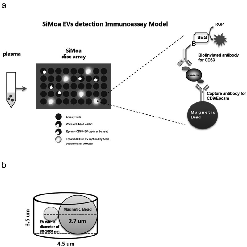
Figure 1.

SiMoa EVs detection immunoassay model.
(A) Samples are incubated with magnetic beads coated with capture CD9 or Epcam antibodies. The bead-EV complexes are incubated sequentially with the biotinylated detector CD63 antibody, SBG and loaded to the SiMoa disc array thereafter. The catalytic reaction of SBG with RGP is restricted in the micro-well. The instrument detects an increasing fluorescent signal if a bead-EV-detector-SBG complex is loaded to the well. (B) The micro-well is large enough for only one magnetic bead binding the EVs/exosomes.
