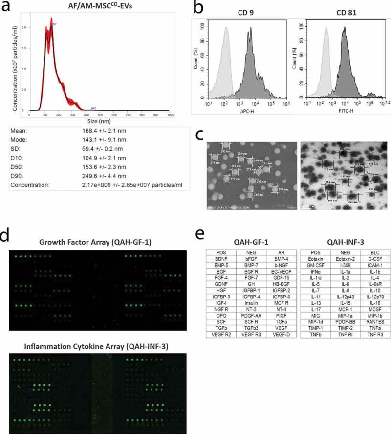
Figure 2.

Characterization of AF/AM-MSCCO-EVs. (a) Particle concentration and distribution of AF/AM-MSCCO-EVs by Nanosight particle tracking system. (b) FACS analysis of AF/AM-MSCCO-EVs to investigate the expression of exosomal markers (CD9 and CD81). (c) Field Emission Scanning Electron Microscope (FE-SEM, left) and Field Emission Transmission Electron Microscope (FE-TEM, right) images of AF/AM-MSCCO-EVs, respectively. (d) Human growth factor and cytokine array of AF/AM-MSCCO-EVs. The upper and lower panel show the scan images of 40 kinds of human growth factors array (QAH-GF-1) and human cytokine array (QAH-INF-3) of AF/AM-MSCCO-EVs, respectively. (E) The left and right panel represent the list of human growth factors and cytokines on each array.
