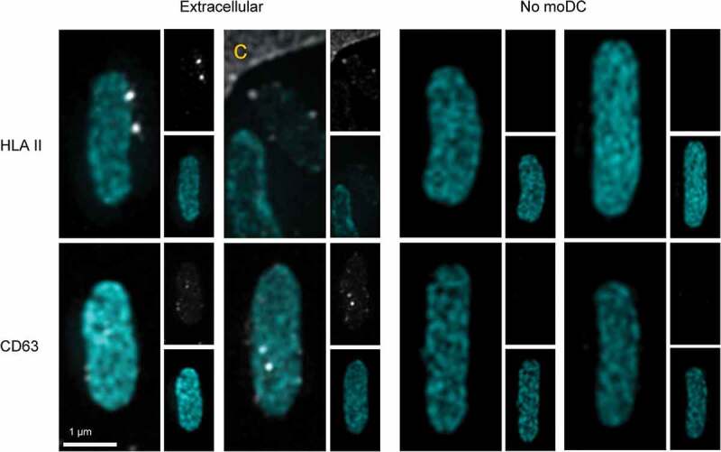
Figure 3.

EV markers on expulsed E. coli.
MoDC were cultured for 16 hour in presence of Cy3B-labelled E. coli. Extracellular E. coli (cyan) that were captured together with moDC were often decorated with HLA II (grey) and CD63 (grey) puncta (left panels), indicating associated EV. Merged pictures show three and two representative examples for HLA II and CD63 respectively. Single channel pictures are displayed in half size prints for comparison. The edge of one MoDC is indicated with c. Examples of control E. coli that had not been incubated with moDC were imaged with identical settings, and the absence of HLA II and CD63 labelling on these EV lacking bacteria demonstrates specificity of the labelling procedure (right panels, indicated with no MoDC).
