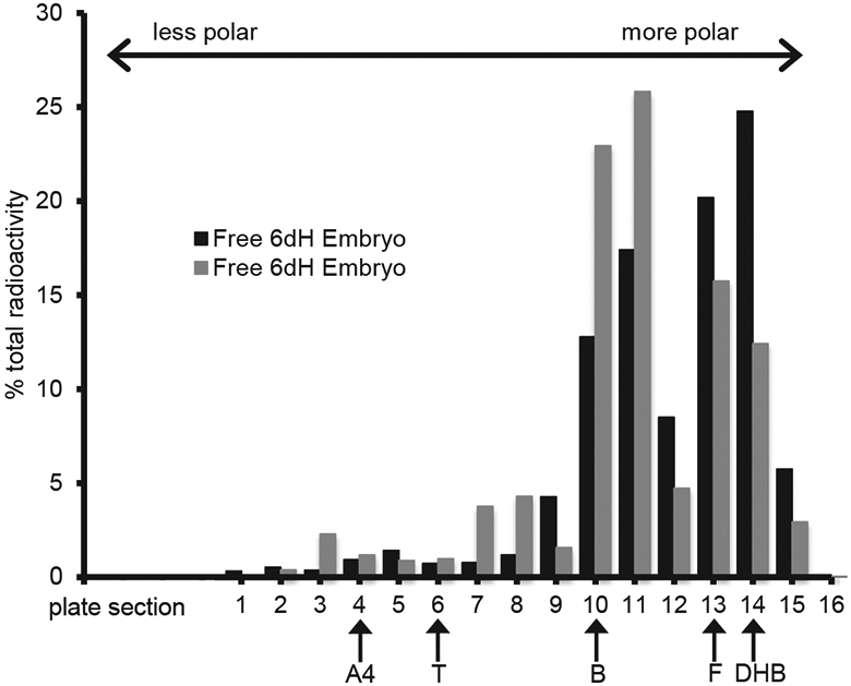
Fig. 3.

Thin layer chromatography based separation of radioactive compounds from two, day 6 high dose apolar (Free) embryo sample fractions. Samples were spotted at section 15 and were run in a solvent system in which free steroids migrate based on their polarity. Plate section 1 represents the point of greatest migration (least polar). Non radioactive steroid standards were run concurrently and their final positions are marked with arrows (A4, androstenedione; T, testosterone; B, corticosterone; F, cortisol; DHB, dihydrocorticosterone).
