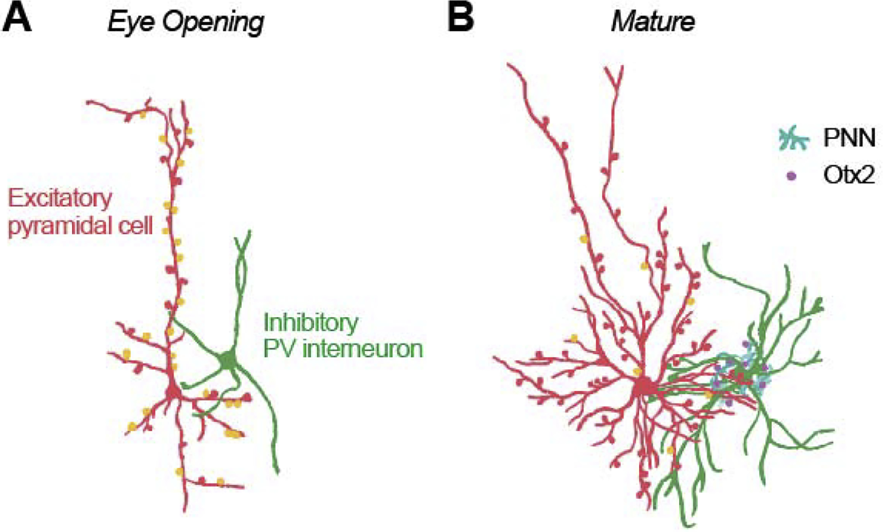
Figure 3.

Illustration of structural changes in development within visual cortex just after eye opening (A) and at the end of the sensitive period (B). Initially, all excitatory neurons (red) appear pyramidal, with a prominent apical dendrite and few small basal dendrites. Later the majority of these neurons gradually sculpt to a stellate morphology [PYR: Conversion to Stellate, ferrets [27]]. Inhibitory PV cells (green) increase in complexity, and the number of inhibitory connections (mostly targeting the soma and proximal dendrites) increases [GABA: GAD65, mouse, [34]; GABA-Aα1, mice, [28]]. Over a slightly longer timeframe there is a decline in the proportion of excitatory synapses that lack AMPA (orange) and are functionally silent [SS: Propn Active; mice [28,39]]. While the number of synapses continues to increase for some time [Synaptic Density, human, [51]], eventually the growth of novel synapses is limited by perineuronal nets (cyan) [PNN, rodents, [44]], which permits the capture of Otx2 (purple).
