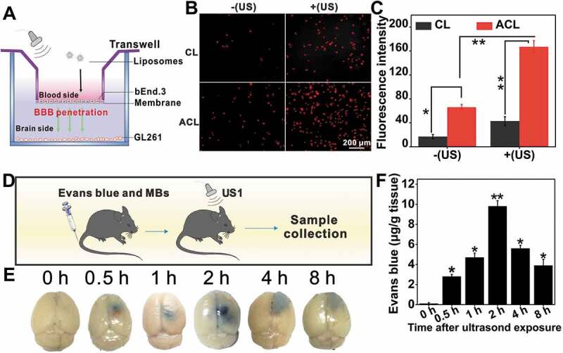
Figure 10.

BBB opening in vitro and in vivo. (A) Illustration of the in vitro BBB model using a Transwell system to evaluate the penetration capability of ultrasound to across the endothelial monolayer. (B) The CL and ACL were added into the upper chambers of the Transwell at a Ce6 concentration of 1 μg/mL for incubation at 37°C for 12 h. Transporting efficiency of liposomes with or without US was measured by Inverted fluorescence microscope at 1 h, scale bar: 200 µm. (C) Quantification of the Ce6 fluorescence intensity with or without US, data are presented as mean ± SD (n = 12 images per group). (D) Illustration of UTMD-induced BBB opening in C57BL/6 mice. (E) Localized leakage of EB at various times after exposed to US. (F) The corresponding quantification of dye content in (E), data are presented as mean ± SD (n = 3 mice per group). *p < 0.05; **p < 0.01.
