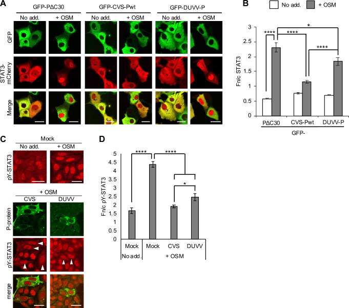
Fig 2. DUVV is defective for STAT3 antagonism compared with RABV.
(A) COS7 cells co-transfected to express the indicated proteins were treated 24 h post-transfection with or without OSM (10 ng/ml, 30 min) before live-cell CLSM. (B) Images such as those shown in A were analysed to calculate the Fn/c for STAT3-mCherry (mean ± SEM; n ≥ 68 cells for each condition; results are from a single assay representative of two independent assays). (C,D) HeLa cells infected with CVS or DUVV (MOI of 1) or mock-infected were treated 48 h post-infection with or without OSM (10 ng/ml, 45 min) before fixation, staining for P-protein (green) and pY-STAT3 (red) and immunofluorescence analysis (C) to determine the Fn/c for pY-STAT3 (D; mean ± SEM, n ≥ 13 cells). Arrowheads indicate cells in which infection was clearly detectable. Scale bars, 30 μm. Statistical analysis used Student’s t test. *, p < 0.05; ****, p < 0.0001.

