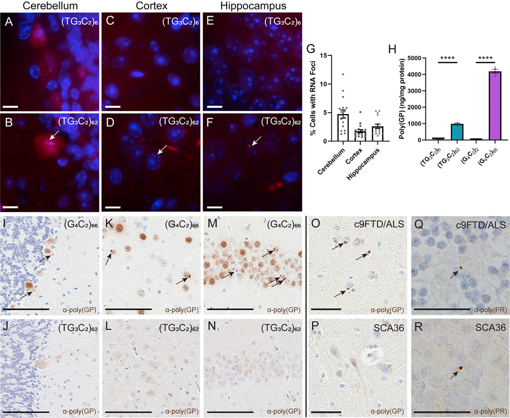
Figure 3. (TG3C2)62 Forms RNA Foci and Undergoes RAN Translation In Vivo.

(A–F) FISH detects sense RNA foci (arrows) in the cerebellum (A and B), cortex (C and D), and hippocampus (E and F) of representative 3- to 4-month-old mice. DAPI marks nuclei in blue. Scale bars: 10 μm.
(G) Distribution of RNA foci in 3- to 4- month-old (TG3C2)62 mice. Similar results are seen at 6 months.
(H) MSD immunoassay to detect poly(GP) in cortical lysates from 3- to 4-month-old mice. Error bars are SEM. n = 5–8 mice per AAV. Samples were run induplicate. ****p ≤ 0.0001 (one-way ANOVA with Tukey’s multiple comparisons test).
(I–N) IHC for poly(GP) in the cerebellum (I and J), cortex (K and L), and hippocampus (M and N) of representative 3- to 4-month-old mice. Arrows mark inclusions. Scale bars: 100 μm.
(O and P) IHC for poly(GP) in human frontal cortex of c9FTD/ALS (O) and SCA36 (P) patients. Arrows mark inclusions. Scale bars: 50 μm.
(Q and R) IHC for poly(PR) in human cerebellar granular cells of c9FTD/ALS (Q) and SCA36 (R) patients. Arrows mark inclusions. Scale bars: 25 μm.
See also Figure S4.
