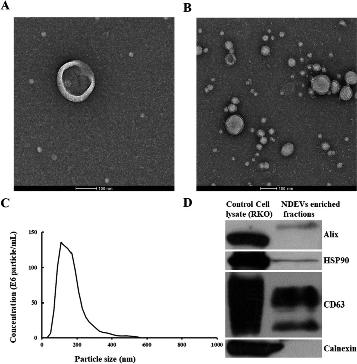
Figure 2.

Plasma NDEVs identified with TEM, NTA, and Western blotting. A single‐plasma NDEV (A) and clusters of plasma NDEVs (B) were measured with TEM. Scale bars equal 100 nm. An NTA plot (C) of size/concentration for plasma NDEVs derived from a patient with AD shows a high concentration. (D) ALIX, HSP90, and CD63 (EV markers) were present and calnexin (a marker for cellular components) was absent in plasma NDEVs enriched fraction samples. Abbreviations: NDEV, neuronally derived extracellular vesicle; TEM, transmission electron microscopy; NTA, nanoparticle tracking analysis; AD, Alzheimer’s disease.
