
Figure 1.
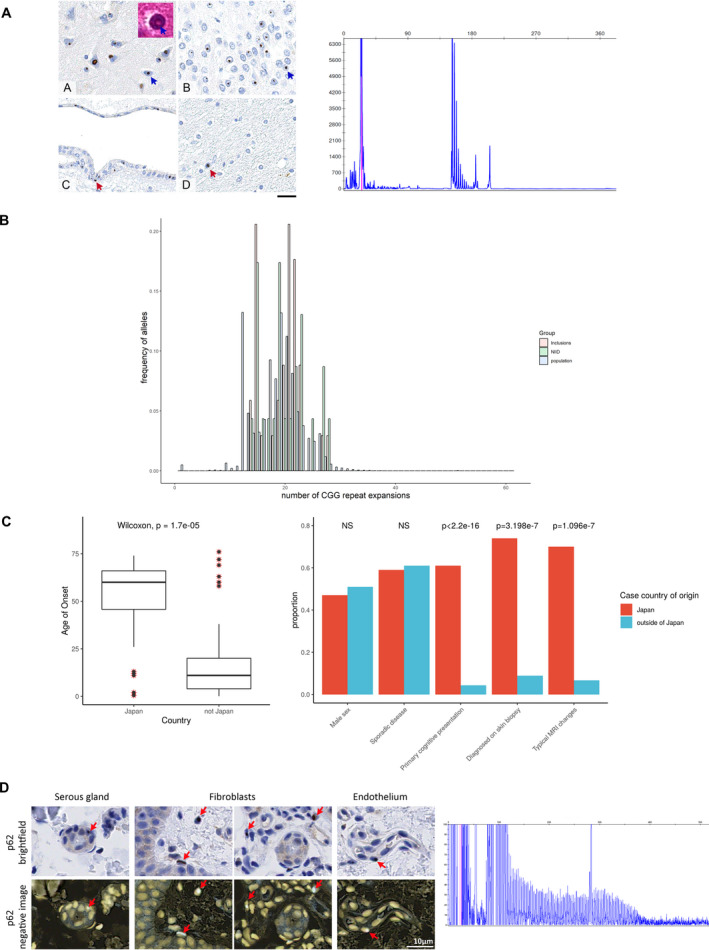
Distribution of NOTCH2NLC repeat expansions. Panel A is that of Patient 1 11 : intranuclear p62 immunoreactive inclusions are present in the majority of the neurons across the neocortex (A, blue arrow), dentate gyrus in the hippocampus (B, blue arrow), deep grey nuclei, brainstem nuclei and cerebellar neurons (not shown). The inclusions are eosinophilic on routine haematoxylin and eosin stained sections (inset in A, blue arrow). The intranuclear inclusions are also frequently seen in the ependymal cells (C, red arrow) but only rarely observed in glial cells (D, red arrow). Scale bar: 20 µm in A‐D. The corresponding electropherogram confirms absence of the repeat expansion within this patient. Panel B shows the histogram distributions of the number of CGG repeats in the population (population) (estimated from ExpansionHunter based on 20,536 participants with neurological presentations enrolled into the 100,000 Genomes Project) compared with cases of neuropathologically confirmed NIID within our samples (NIID) and cases with evidence of pathological intranuclear inclusions (inclusions). Panel C summarizes the comparison of clinical characteristics between cases of NIID described within and outside of Japan. Panel D shows brightfield‐positive and brightfield‐negative images for p62 immunoreactivity in the skin biopsy of the patient identified from 100,000 Genomes Project (Case 12). The corresponding electropherogram infers presence of a repeat expansion as seen by the typical sawtooth pattern.