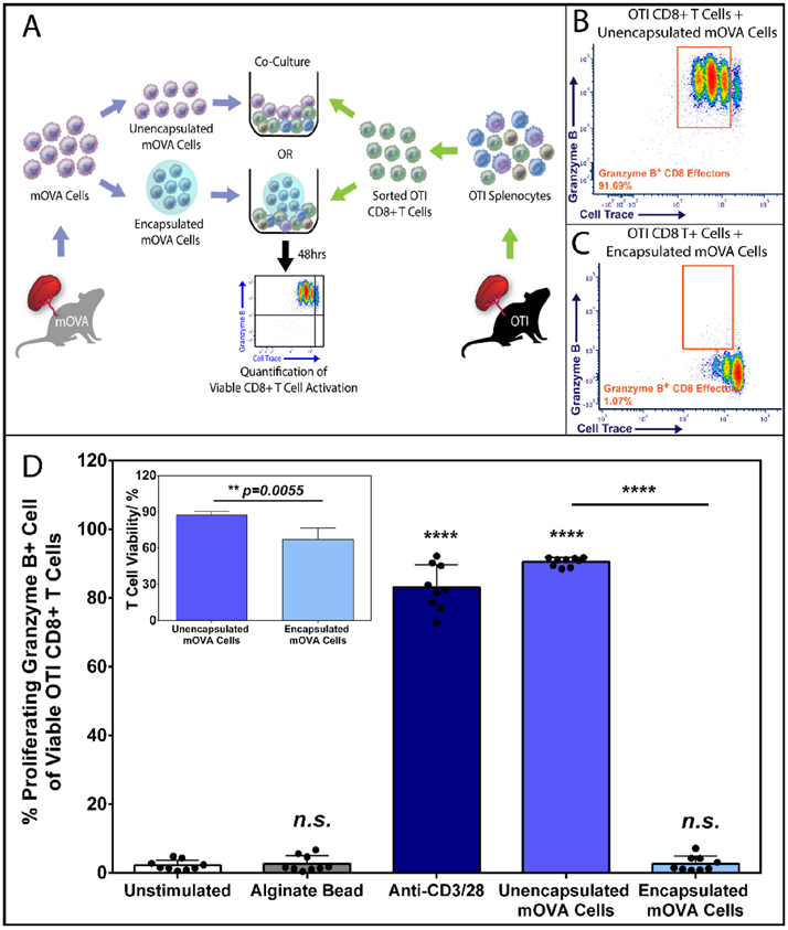
Figure 2. Alginate Encapsulation Blocks Cell-Contact Dependent Direct CD8+ T Cell Antigen Recognition and Activation.

(A) Schematic overview of the 48hr coculture experiment co-incubating sorted OTI CD8+ T cells (see Figure S1 for sorting) with unencapsulated or encapsulated mOVA stimulatory splenocytes at 1:1 ratio. Antigen-specific OTI CD8+ T cell activation was quantified by the % of proliferating (Cell Trace® Violet labeled) and granzyme B + CD8+ T effector cells (see Figure S2 for gating) via flow cytometry analysis. Representative FCM data of OTI CD8+ T cell activation by unencapsulated (B) or alginate encapsulated (C) mOVA cells after a 48-hour stimulation. (D) Summary of the frequency of proliferating granzyme B+ CD8+ T effectors in response to designated stimuli (x-axis). Inset: CD8+ T cell viability after 48 h incubation with unencapsulated or encapsulated mOVA cells. Bars indicate the average of individual data points (N=3; n=9) with standard deviation. Statistical significance was determined as ****p < 0.0001 and n.s. = not significant via Tukey’s test when compared with the unstimulated control group.
