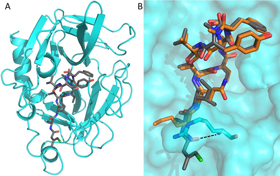
Figure 3.
A Cartoon representation of PPE (cyan) in complex with 1 (shown as grey sticks). B Comparison of 1 (grey) and 4 (PDB: 4GVU; orange) binding to PPE (cyan surface). The side chains of PPE residues Ser216 and Arg217 are shown as sticks. The additional hydrogen bond found in the PPE-1 complex structure is indicated by a dashed line.

