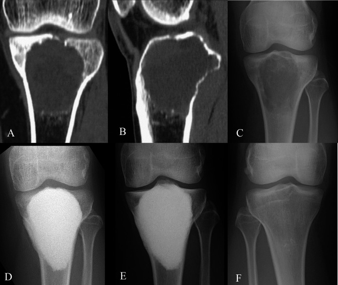
Fig. 1.
Figs. 1-A through 1-F Preoperative CT and anteroposterior radiographs showing change in the knee joint of a 31-year-old woman with GCTB at the proximal aspect of the left tibia (Case 12, Table I). Fig. 1-A Coronal plane of the lesion with partial subchondral bone loss. Fig. 1-B Sagittal plane of the lesion with partial subchondral bone loss. Fig. 1-C Preoperative state. Fig. 1-D State at 3 months postoperatively. Fig. 1-E State at about 12 years postoperatively. Fig. 1-F State of the nonoperative contralateral knee at about 12 years postoperatively.

