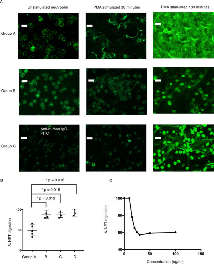
Fig. 3. Binding of humAbs to neutrophils and NET.
a Representative mAb binding to neutrophils and NET from group A, B and C. Group D mAbs. The antibody binding to NET was detected by FITC-conjugated anti-human IgG while NET was visualised by DAPI, bar length = 25 μm. Neutrophils were either unstimulated or stimulated with 100 ng/ml PMA for 30 or 180 min before staining. Groups A and B bound to neutrophil cytoplasm at 10 μg/ml as shown in the figure but did not bind to unstimulated neutrophil cytoplasm at 1 μg/ml. In total, 12/46 mAbs were positive for NET binding. b Percentage of NET digestion was measured in the presence of group A, B, C and D mAbs (n = 15) and displayed as a dot plot, repeated for three times. Data represent mean ± SEM, Error bars indicate mean with 95% CI and Mann–Whitney U was performed. c Group A mAbs protect NET from nuclease digestion in a dose-dependent manner, ranging from 1 to 100 μg/ml. The graph is representative of one group A mAb (557A3), n = 15.

