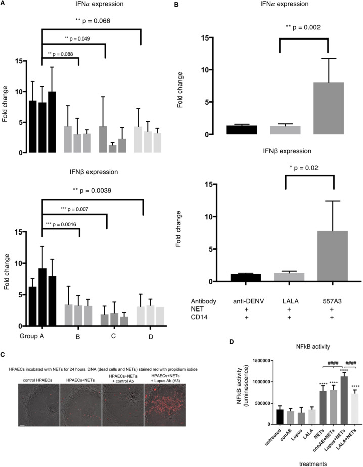
Fig. 4. Group A antibodies enhance type I IFN and NF-kB.
a Monocytes were co-incubated with NET and three humAbs from each group (n = 12). Type I IFN expression was then measured by qPCR and fold change were calculated against negative control (anti-DENV antibody). Error bars indicate mean of three repeats with 95% CI, two-way ANOVA. b Type I IFN mRNA levels are measured by incubating NET, monocyte and group A humAb-557A3 (dark grey) or 557A3-LALA (light grey). Error bars indicate mean with 95%CI Student t-test, n = 4. c Significantly more NET stayed on HPAECs after 24 h when co-incubated with group A humAb-557A3. HPAECs were incubated with NETs for 24 h in the presence of control and 557A3 before staining with propidium iodide. The addition of group A antibody markedly attenuated degradation of NETs (red, last image on the right). Bar = 50 µm. d HPAECs infected with AdNFkB-luc were incubated with NETs in the presence or absence of group A antibody 557A3. Following overnight incubation, NF-kB activity was measured in a luciferase reporter assay. ****P < 0.0001, comparisons with untreated control; ####P < 0.0001, comparisons, as indicated. ANOVA with Tukey post-test, n = 4.

