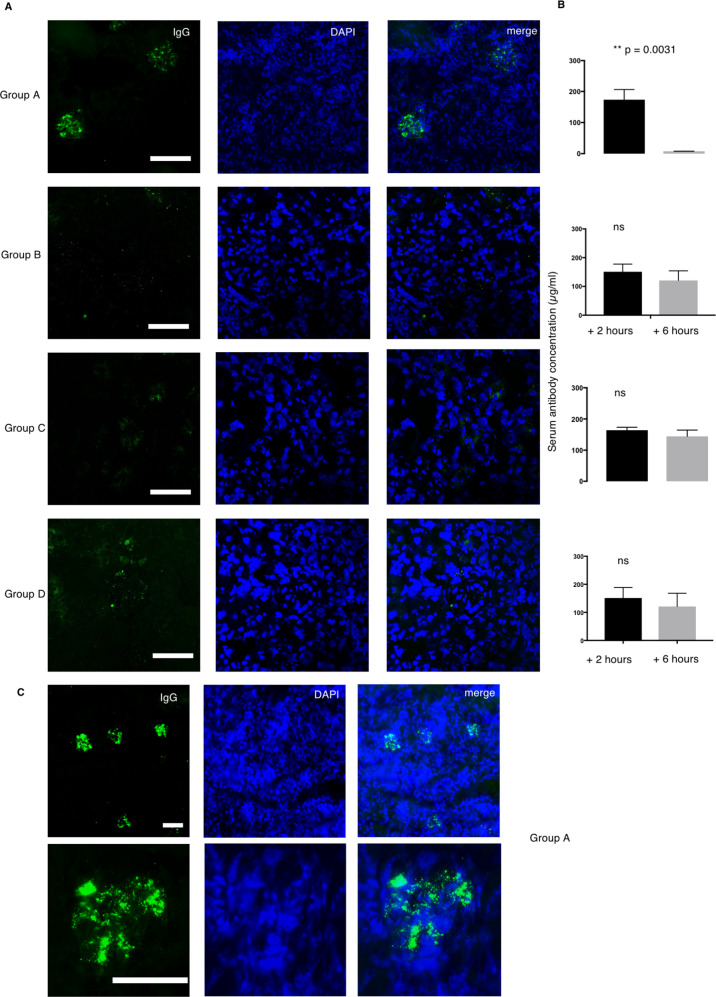
Fig. 5. Group A antibody deposit in the kidney of nephritic mice.
a Two humAbs from Group A, B, C and D (n = 8) were intraperitoneally injected to the nephritic mice. After six hours the mice were sacrificed to collect serum and kidney sample. IgG deposition was studied by staining the acetone fixed kidney with FITC-conjugated anti-human IgG (green) and DAPI (blue). b Serum human IgG level was measured at 0 and 6 h after humAb injection. Data represent mean ± SEM. Error bars indicate mean with 95% CI. n = 3. Student t-test. c The deposition was observed in multiple glomeruli seen at ×10 magnification and deposition pattern was visualised at ×40 magnification.

