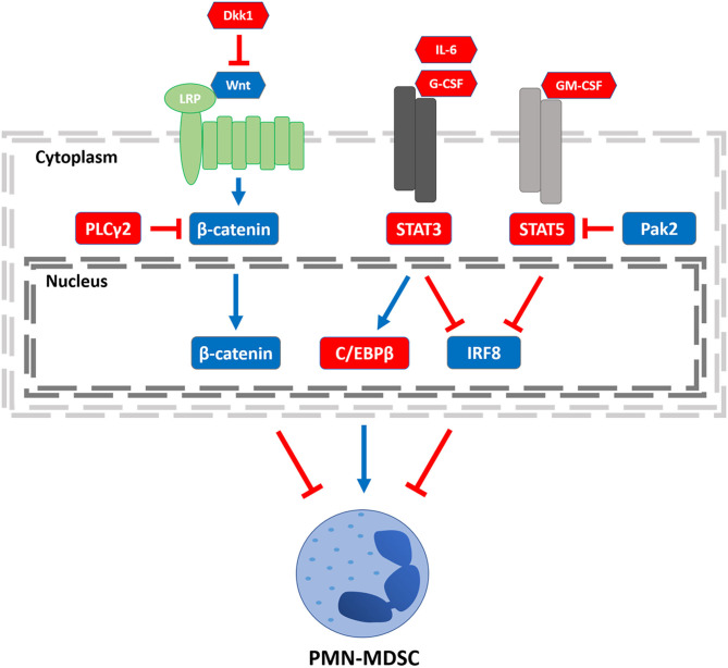
Figure 1.
Transcriptional regulators of PMN-MDSCs. Three regulatory axes of PMN-MDSC development or function are depicted. Left to right: PLCγ2 and Dkk1 have been shown to decrease β-catenin signaling, increasing PMN-MDSC burden. STAT3 signaling can be activated by stromal- or tumor-derived factors such as IL-6 or G-CSF, enhancing C/EBPβ expression. STAT3 activation can also inhibit IRF8 expression. Both axes lead to an increase in PMN-MDSCs, although it remains to be determined whether there is crosstalk between the C/EBPβ and IRF8 pathways. Stromal- or tumor-derived GM-CSF engagement leads to STAT5 activation, which inhibits IRF8 expression, an effect that can be overcome by Pak2-mediated inhibition of STAT5. Legend: nodes shown in red or blue enhance or block PMN-MDSCs, respectively; arrows shown in red or blue inhibit or activate its downstream target, respectively.

