Figure 2.
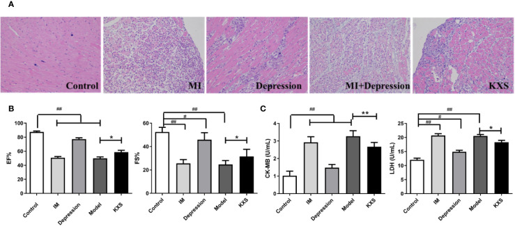
The cardioprotective effect of KXS. Rats were randomly assigned into five groups: control, MI, depression, model and KXS. After experiment, the myocardial injury was determined by HE staining, echocardiography (EF and FS) and cardiac marker enzymes (CK-MB and LDH). (A) Representative HE staining of myocardial tissue (×200). (B) The EF and FS changes in groups. (C) The CK-MB and LDH changes in groups. Compared with control group, #P < 0.05, ##P < 0.01; Compared with model group, *P < 0.05, **P < 0.01. EF, ejection fraction; FS, fractional shortening; CK-MB, serum creatine kinase MB; LDH, lactate dehydrogenase; ISO, isopropyl adrenaline; Control, normal rats; IM, injected ISO; Depression, chronic mild stress rats; Model, depression with ISO; KXS, KXS+ depression with ISO.

