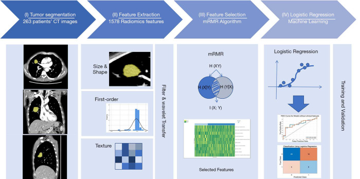
Figure 1.
The study workflow diagram. (I) Tumor segmentation: the tumor was three-dimensionally semi-automatically segmented in chest CT images. (II) Feature extraction: 1,672 radiomic features were automatically extracted from the segmented tumor volume. (III) Feature selection: radiomic features were selected using the minimum redundancy maximum relevance algorithm. (IV) Logistic regression: models were trained and tested to determine their classification performance

