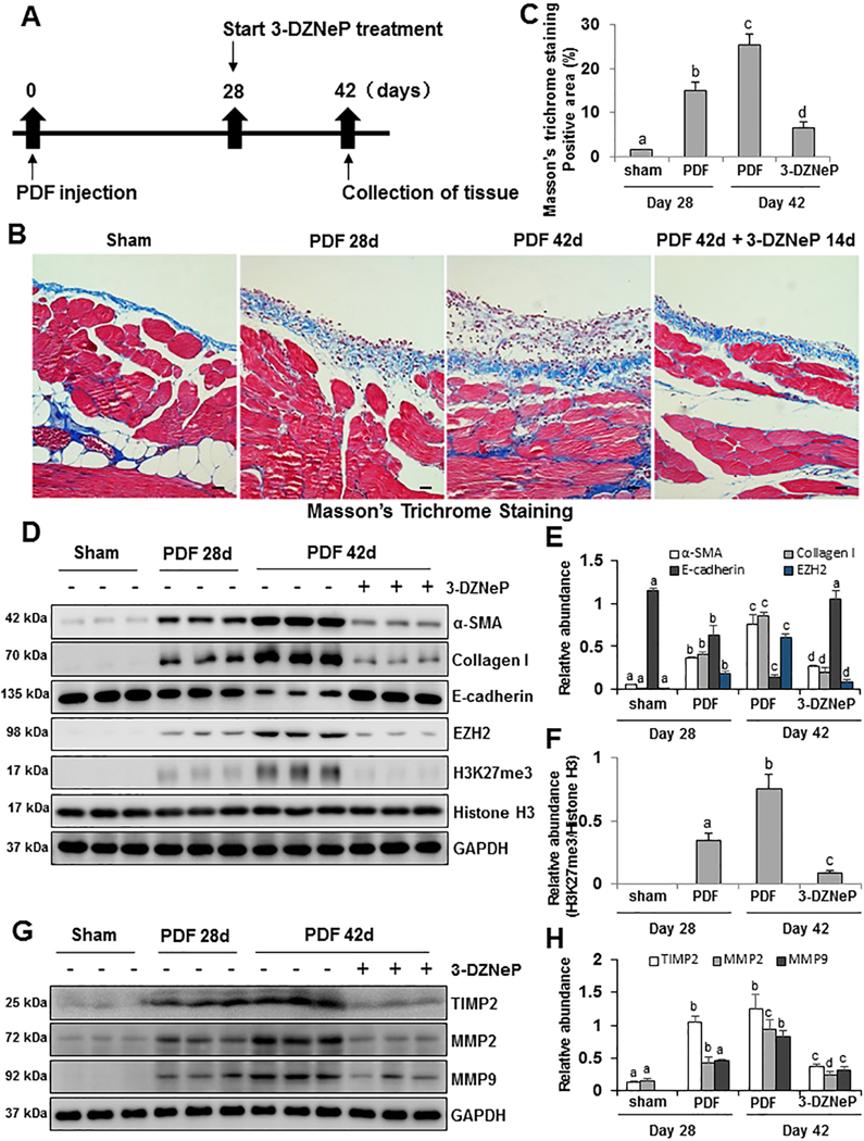
Figure 5. Delayed treatment with 3-DZNeP attenuates and reverses progression of peritoneal fibrosis and suppresses EMT.
(A) Schematic of the experimental design for delayed treatment with 3-DZNeP. To investigate the therapeutic effect of 3-DZNeP on peritoneal fibrosis mice models, 3-DZNeP was administered starting at 28 days after injection with 4.25% high glucose dialysate and then given daily for 14 days. At 42 days, animals were euthanized for collection of peritoneum. (B) Photomicrographs (200X) show Masson trichrome staining of the peritoneum in each group. (C) The graph shows the positive area of the Masson-positive submesothelial area (blue) from ten random fields of six mice peritoneal samples. (D) Peritoneum tissue lysates were subjected to immunoblot analysis with specific antibodies against α-SMA, Collagen I, E-cadherin, EZH2, H3K27me3, Histone H3 or GAPDH. (E) Expression levels of α-SMA, Collagen I, E-cadherin and EZH2 were quantified by densitometry and normalized with GAPDH. (F) Expression level of H3K27me3 was quantified by densitometry and normalized with Histone H3. (G) Peritoneum tissue lysates were prepared and subjected to immunoblot analysis with antibodies against TIMP2, MMP2, MMP9, or GAPDH. (H) Expression levels of TIMP2, MMP2 and MMP9 were quantified by densitometry and normalized with GAPDH. Data are represented as the mean ± S.E.M (n = 6). Means with different superscript letters are significantly different from one another (P< 0.05). All scale bars = 20 μm.

