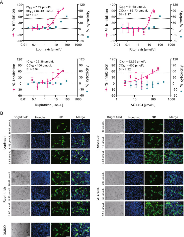
Fig. 2.
In vitro inhibition of viral protease inhibitors against SARS-CoV-2 in Huh7 cells. Huh7 cells were infected with SARS-CoV-2 at an MOI of 0.1 in the treatment of different concentrations of the indicated compounds. At 48 h p.i., cell supernatants were collected and cells were fixed. A The viral yield in the cell supernatant was quantified by qRT-PCR. Cytotoxicity of these drugs to Vero E6 cells was measured by CCK-8 assays. The left and right Y-axis of the graphs represent mean % inhibition of virus yield and % cytotoxicity of the drugs, respectively. The experiments were done in triplicates. B Fixed cells were subjected to IFA by employing anti-NP rabbit sera. The nuclei were stained with Hoechst dye. Bars, 100 μm.

