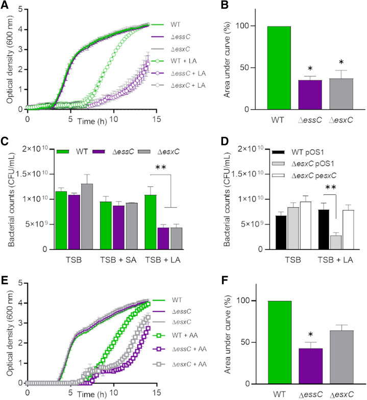
Figure 1.
Enhanced S. aureus growth inhibition by antimicrobial fatty acids in esxC and essC mutants. (A) S. aureus WT USA300, ΔessC, and ΔesxC were grown in TSB or TSB supplemented with 80 µM linoleic (LA). Means ± standard error of the mean (SEM) are shown. n = 4. (B) The area under the curve (AUC) of biological replicates grown in TSB + LA in (A) were calculated and presented as % relative to the WT. Means ± SEM are shown. *Indicates P < 0.05 using a Kruskal–Wallis test with Dunn's multiple comparisons test. (C) After 14 h growth in TSB or TSB supplemented with 80 µM LA or stearic acid (SA), bacteria were serially diluted, and CFU were determined. Mean values are presented, and the error bars represent SEM. n = 3, **indicates P < 0.01 using one-way ANOVA with Dunnett’s test. (D) USA300 WT with the empty pOS1 plasmid (WT pOS1) and USA300 JE2 esxC mutant with either pOS1 (ΔesxC pOS1) or pOS1-esxC (ΔesxC pOS1-esxC) were grown in TSB or TSB + 80 µM LA as described in (A) followed by CFU estimation. Mean values are shown; error bars represent SEM. n = 5, **indicates P < 0.01 using one-way ANOVA with Dunnett’s test. (E) S. aureus WT USA300, ΔessC, and ΔesxC were grown in TSB or TSB supplemented with 80 µM arachidonic acid (AA). Means ± SEM are shown, n = 3. (F) AUCs of biological replicates grown in TSB + AA in (E) were calculated and presented as % relative to the WT. Means ± SEM are shown. *Indicates P < 0.05 using a Kruskal–Wallis test with Dunn's multiple comparisons test.

