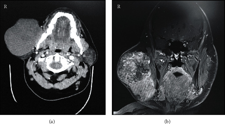
Figure 2.

Preoperative radiographic examination of the right preauricular swelling. (a) Contrast-enhanced computed tomography axial section at the level of mandibular teeth shows a well-defined mass lesion in the superficial lobe of the right parotid gland, without any underlying bony erosion and normal-appearing pharynx, larynx, and parapharyngeal spaces. (b) Magnetic resonance imaging coronal section along the posterior border of the mandible shows a large, heterogeneous, well-demarcated solid mass lesion within the right parotid superficial lobe and measuring 10 × 7 × 8 cm at maximum dimensions.
