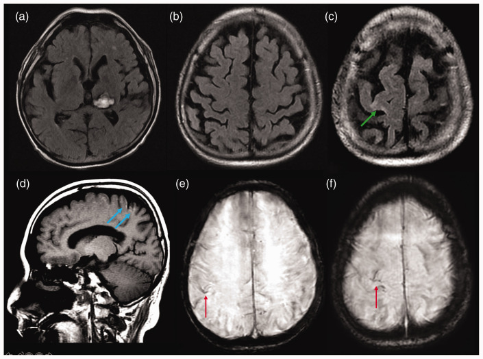
Figure 3.
Follow-up brain magnetic resonance imaging with conventional imaging using axial FLAIR ((a), (b) and (c)), sagittal T1 (d) and axial SWI ((e )and (f)), showing partial reabsorption of the subarachnoid haemorrhage, foci of cortical laminar necrosis (blue arrows), gliosis in the right prefrontal cortex (green arrow), superficial siderosis (red arrows) and resolution of both cortical and subarachnoid contrast-enhancement, with no additional signs of acute ischaemia.

