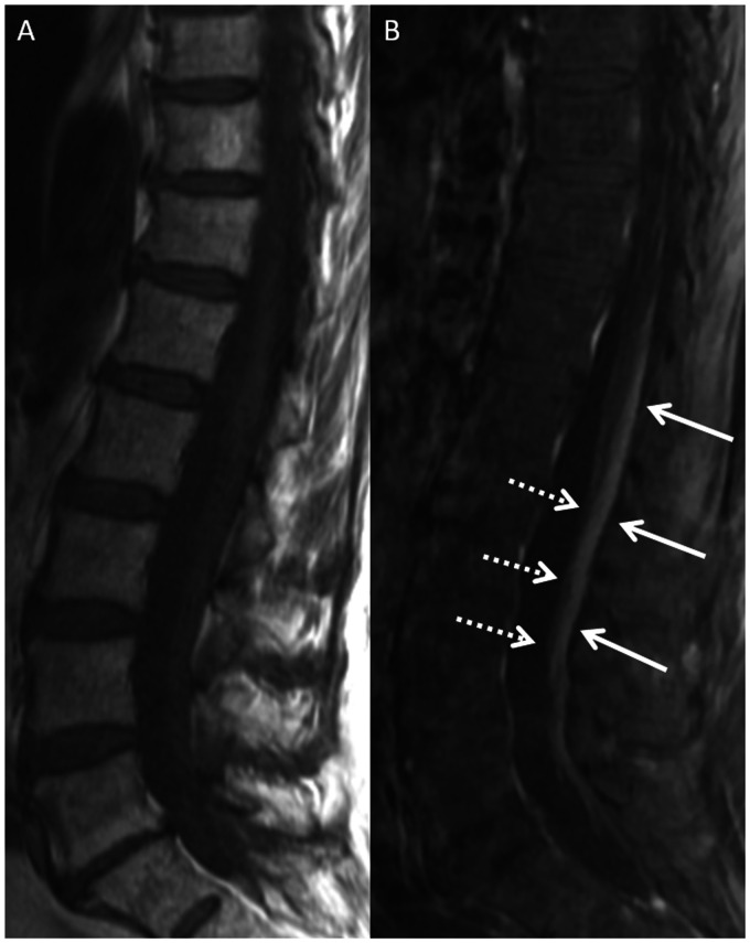
Figure 1.
Magnetic resonance imaging (MRI) of the lumbar spine in a 69-year-old female with paraneoplastic polyneuropathy. Sagittal T1-weighted (T1W) pre-contrast (a) and post-contrast (b) images show smooth enhancement of the dorsal cauda equina nerve roots (b, solid arrows). There is no appreciable enhancement of the ventral nerve roots (b, dashed arrows).

