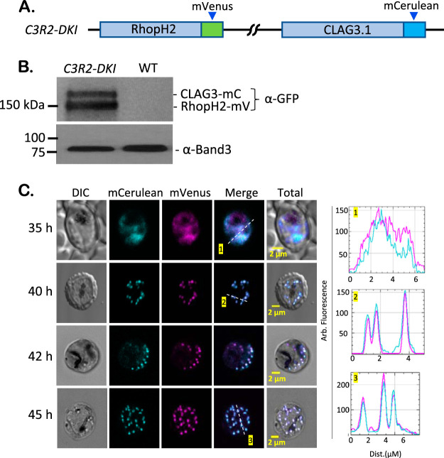
FIG 3.
Strict association between RhopH2 and CLAG3 revealed by imaging tandem-labeled cells. (A) Ribbon diagram showing tandem labeling of RhopH2 and Clag3h through sequential CRISPR-Cas9 transfection to produce the C3R2-DKI clone. (B) Immunoblot of total cell lysates from indicated parasites, probed with anti-GFP antibody. Two bands are detected in C3R2-DKI, consistent with tagging of both CLAG3 and RhopH2. Loading control, Band3. (C) Live-cell images of C3R2-DKI-infected cells at the indicated time points after invasion. RhopH2 and CLAG3 colocalize within trophozoites (top row) and within rhoptries of mature schizonts (other rows). Right panels, corresponding line scans. The results are representative of >80 cells imaged from at least eight experiments.

