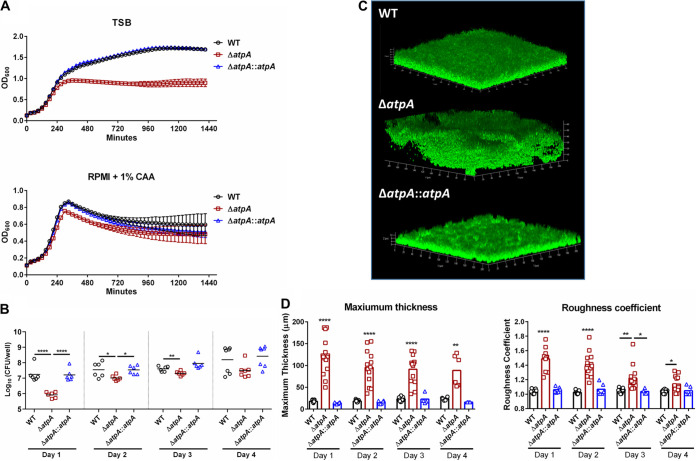
FIG 1.
S. aureus ΔatpA biofilm displays early growth defects and altered structure. (A and B) The growth of S. aureus WT, ΔatpA, and ΔatpA::atpA was characterized by OD600 in tryptic soy broth (TSB) or RPMI-1640 supplemented with 1% Casamino Acids (CAA; mean ± SD of one representative experiment; n = 6 biological replicates) (A) and CFU of in vitro biofilm at various stages of development (mean combined from 2 independent experiments; n = 6 biological replicates) (B). (C) Representative three-dimensional (3D) images of 4-day-old biofilm acquired using confocal laser scanning microscopy. (D) Maximum thickness and roughness coefficient measurements were calculated by Comstat 2 analysis (mean combined from 1 to 4 independent experiments; n = 3 to 15 biological replicates). Significant differences are denoted by asterisks (*, P < 0.05, **, P < 0.01, and ****, P < 0.0001; one-way ANOVA with Tukey’s multiple-comparison test).

