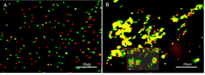
FIG 3.
(A) Confocal scanning laser microscope image of protein binding assay showing the dispersal of green and red microspheres following preincubation with a nonbinding protein (i.e., bovine serum albumin [negative control]). Note that the majority of beads with different fluorescence are observed separately (i.e., as red or green only), with a few overlapping signals (i.e., yellow) due to random positional overlay. (B) Coalescence of green microspheres incubated with construct 1 (i.e., anammox biofilm S-layer protein amino acids 1254 to 1338) and red microspheres incubated with construct 2 (i.e., amino acids 1354 to 1439). (Inset) Three-dimensional side view of coalesced beads, demonstrating that binding derives from protein interactions and not the overlapping of beads. The samples were prepared in 50 mM MES–5 mM CaCl2, pH 6.0.

