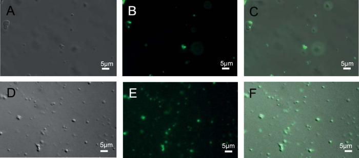
FIG 5.
Bright-field (A and D), epifluorescence (B and E), and superimposed (C and F) micrographs of liquid droplets of an anammox biofilm surface protein construct (amino acids 1254 to 1338) (100 μM) in an aqueous solution of 20 mM Tris (pH 7.5)–125 mM NaCl–2 mM DTT, 4°C, and polyethylene glycol (20% [wt/vol]) following the addition of 0.5-μm carboxylate-modified latex microspheres (green) (B and C) and green fluorescence protein-labeled Pseudomonas aeruginosa cells (E and F) showing the wetting and protein droplet-mediated coalescence of microspheres and bacterial cells, respectively.

