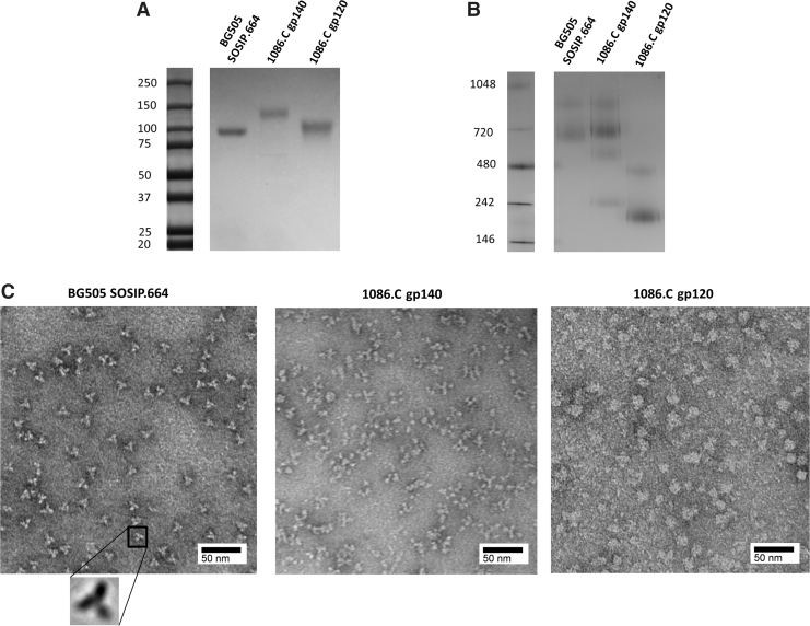
FIG. 1.
NS-EM and PAGE analysis of HIV-1 Env vaccine candidates. (A) Western blot analysis of BG505 SOSIP.664, 1086.C gp140, and 1086.C gp120 Envs under reducing condition. (B) BN-PAGE analysis of BG505 SOSIP.664, 1086.C gp140, and gp120 (2 μg of each protein). The molecular weight of marker (M) proteins are indicated (B, C). (C) NS-EM analysis of BG505 SOSIP.664, 1086.C gp140, and 1086.C gp120 Envs stained with 7.5% uranyl formate. Electron microscopy was performed using Tecnai G2 Spirit BioTWIN at 80 kV. Scale bar, 50 nm. BN-PAGE, blue native–polyacrylamide gel electrophoresis; Env, envelope glycoprotein; NS-EM, negative staining electron microscopy.

