Fig. 5.

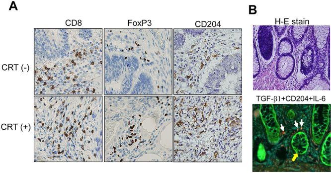
Tumor-infiltrating immune cell characterization using IHC and immuno-fluorescence staining. (A) IHC staining against CD8, Foxp3 and CD204. CRT(−) and CRT(+) specimens were derived from casees REC011 and REC007, respectively. (B) H-E staining and immuno-fluorescent staining for TGF-beta1 (green), IL-6 (blue) and CD204 (red) using the Opal 4-color kit. The white arrows show CD204+ macrophages producing IL-6. The yellow arrows show TGF-beta1+ rectal cancer cells. The specimen was derived from case REC006 with CRT(+) which showed the upregulation of IL-6 gene in a quantitative PCR. Magnification x400.
