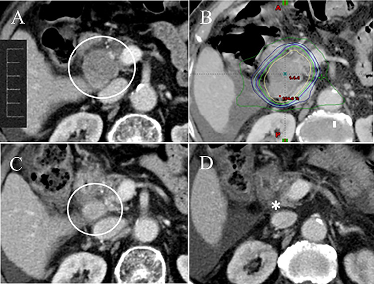
Fig. 1.

A 70-year-old female with HCC treated with TACE. (A) Abdominal CT scan showed an enlarged lymph node metastasis in the retropanceratic space with duodenal obstruction (circle). (B) Abdominal CT with dose distribution curves of SBRT delivered with IMRT. The innermost line was the 100% isodose line (yellow line). SBRT was administered with 45 Gy in 6 fractions (BED, 78.8 Gy10). (C) Abdominal CT at 2 months following the completion of SBRT showed a reduction of the metastatic node size (circle), and the RECIST response rate was partial response (PR). The duodenal obstruction was relieved. (D) The metastatic node disappeared on routine follow-up CT at 9 months following the completion of SBRT (asterisk).
