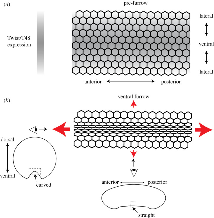
Figure 1.
The mechanical pattern and pattern of apical constriction during mesoderm invagination. (a) The gradient in the expression of the transcription factor Twist and its target T48 before apical constriction and furrow formation. Expression is highest at the ventral midline and drops towards the sides of the embryo. (b) Apical constriction is coordinated so that 4–6 cells at the ventral midline constrict the most and there is a gradient of constriction extending towards the sides of the embryo. Red arrows indicate tension. Eyes indicate different views of the furrow, a dorsal–ventral cross section (left) and mid-sagittal section (bottom).

