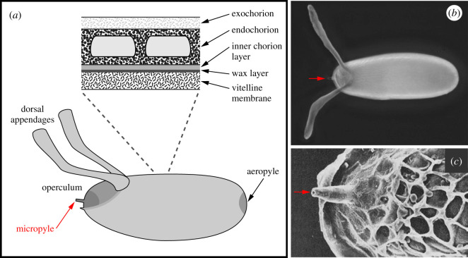
Figure 1.
Structure of the Drosophila eggshell. In all images, anterior is to the left. (a) Illustration highlighting the layers and specialized regions of the eggshell. Illustration is adapted from reference [3]. (b) Light micrograph of the eggshell, dorsal view. The red arrow points to the micropyle. (c) Scanning electron micrograph of an anterior region of the eggshell, dorsal view. The red arrow points to the micropyle. Image is reprinted from reference [4] with permission from Journal of Cell Science.

