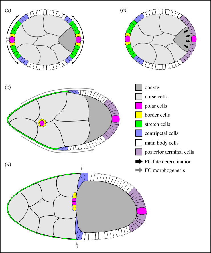
Figure 2.
Illustrations of select stages of egg chamber development. In all images, anterior is to the left. (a) Signals from the polar cells induce the nearby follicle cells (FCs) to adopt a variety of cell fates. (b) A second signal from the oocyte creates a new cell fate at the egg chamber's posterior. (a,b) Both signalling events occur before stage 6. (c) A combination of oocyte growth and epithelial cell shape changes/migrations that occur during stage 9 brings the bulk of the follicle cells into contact with the oocyte. (d) Migration of the centripetal cells between the oocyte and nurse cells late in stage 10 allows them to meet up with the border and polar cells to form a continuous epithelium around the oocyte's anterior. Illustrations are adapted from references [9,10].

