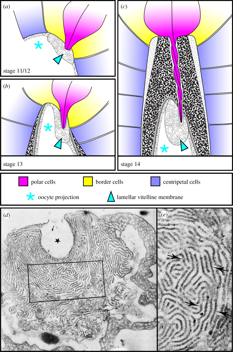
Figure 3.
Micropyle morphogenesis and structure. In all images, anterior is to the top. (a–c) Illustrations of micropyle formation adapted from references [16,17]. (a) At stage 11/12, the centripetal and border cells are secreting the spongy and lamellar vitelline membrane, respectively, and the polar cells have extended stout protrusions. (b) At stage 13, the centripetal cells have deformed the oocyte and are beginning to secrete the chorion layers. (c) At stage 14, micropyle morphogenesis is complete, and the polar cell protrusions twist around one another inside the channel. (d,e) Transmission electron micrographs of a thin section taken through the lamellar vitelline membrane within the mature micropyle of an unfertilized egg. (d) The maze-like organization of the lamellae and the ‘pocket’ (asterisk) made by the tips of the polar cell protrusions are both visible. (e) Blowup of the boxed region in (d). Arrows point to individual lamellae. Images are reproduced from reference [17], © Canadian Science Publishing or its licensors.

