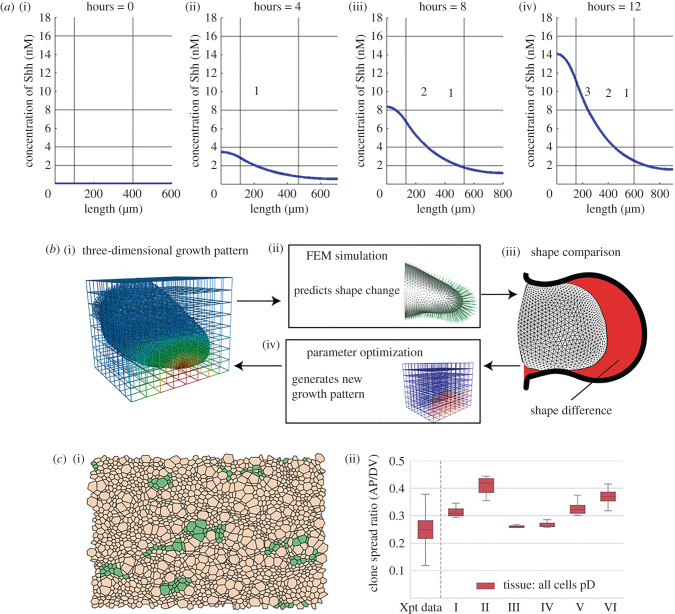
Figure 3.
Modelling Shh in patterning and growth. (a) Simulation of chick wing digit specification by a Shh gradient in a uniformly linearly growing domain. Vertical black lines delineate the domain into three sections; horizontal black lines illustrate the thresholds required for each digit identity. Reproduced with permission from [73]. (b) Schematic illustrating how parameters in a three-dimensional model of the developing limb are fitted by comparing simulated shape dynamics to in vivo data. FEM, finite element model. Reproduced with permission from [74]. (c) Simulation of progenitor proliferation and differentiation in the developing neuroepithelium. (i) Simulated clones reveal a shape bias in the DV direction (dorsal progenitor cells are brown, green represents clones of these dorsal progenitor cells). (ii) Comparison of clone shape bias for six different parameter sets with in vivo data (Xpt data). pD, dorsal progenitor. Reproduced with permission from [75].

