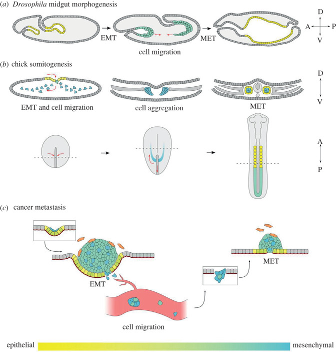
Figure 2.
Successive rounds of EMT and MET take place in development and cancer. (a,b) Several developmental examples exist in which both EMT and MET of the same cell population can be studied. (a) Drosophila embryonic midgut morphogenesis. Midgut cells at both the anterior and posterior of the embryo are initially epithelial (yellow). These cells then undergo a partial EMT (green), which facilitates their collective migration through the embryo. The majority of these migrating cells subsequently undergo MET to form a contiguous midgut epithelium. The compass indicates the anterior–posterior and dorsal–ventral axes. (b) Chick somitogenesis. The upper row of diagrams shows transverse cross sections. Sectioned regions are indicated by dotted lines on the lower row of diagrams, which depict a dorsal view of the embryo. Epithelial cells (yellow) at the primitive streak first undergo EMT to form mesenchymal mesoderm progenitors (blue). The paraxial mesoderm cells (blue) then undergo migration towards the anterior of the embryo to form the presomitic mesoderm (green). The presomitic mesoderm undergoes MET during segmentation to form somites. Mature somites consist of epithelial cells (yellow) enclosing mesenchymal cells (blue). (c) EMT and MET underlie distinct stages of tumour metastasis. Cancerous epithelial cells proliferate and can undergo partial or full EMT to form heterogeneous tumours (coloured) within an otherwise non-cancerous tissue (grey). Tumours also recruit other, non-cancerous cells to facilitate growth and invasion, such as tumour-associated fibroblasts (orange). Migratory cancer cells can escape the tissue via individual or collective migration and enter the circulatory system as individual cells or as clusters, dependent on EMT state. Metastatic colonization of secondary sites requires re-epithelialization of the tumour cells through MET, resulting in the formation of secondary tumours.

