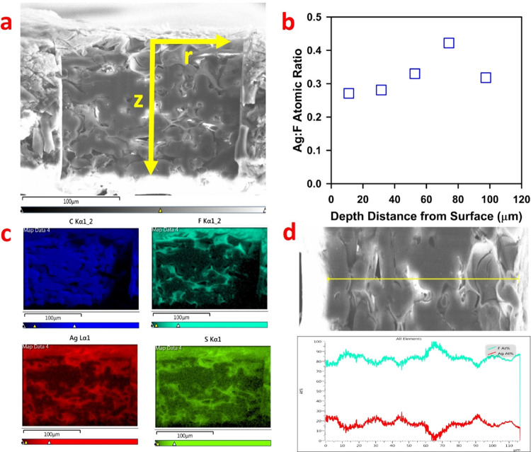
Figure 6.
Characterization of the cross section of the CysM-PAA-PVDF membrane using the FIB instrument to assess the elemental composition after adsorption of Ag as a model compound. The FIB was used to prepare the entire cross section (∼120 μm) with an ion beam (2.5–6 nm), ensuring minimum damage of the sample. (a) Sample of the whole membrane cross section. The smooth area in the center was removed by the FIB, and the elemental composition is assessed in both the z and r direction. (b) Ag to F atomic ratio in different depths of the membrane, confirming an almost even adsorption of Ag+ cations across the whole cross section (i.e., the whole pore) of the membrane. F is used as a standard as it is homogeneously distributed in the PVDF membrane. (c) Distribution profile of atomic C, F, Ag, and S across the entire cross section from the top surface is demonstrated. Ag (red) and S (green) are almost evenly distributed confirming that all the thiol (−SH) sites are utilized. (d) Line scan data of F and Ag atomic percentage in the r direction at a distance of 53 μm from the top surface.

