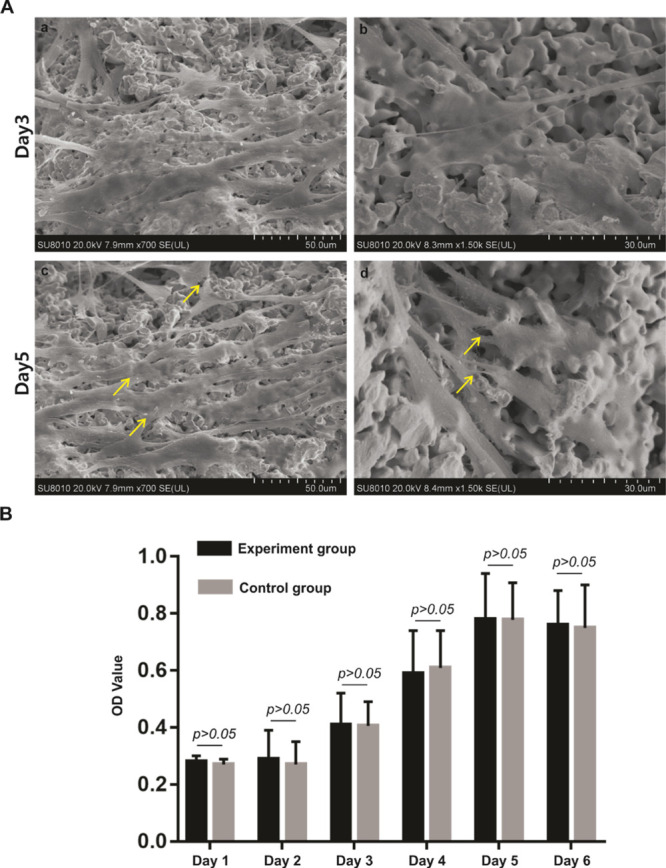
Figure 2.

Cell growth on a novel porous tantalum scaffold and cytotoxicity evaluation of porous tantalum scaffold. (A) SEM micrographs of UC-MSCs on the porous tantalum scaffold at different time points. UC-MSCs on the porous tantalum scaffold on day 3 (a, scale bar = 50 μm, b, scale bar = 30 μm). UC-MSCs on the porous tantalum scaffold on day 5 (a, scale bar = 50 μm, b, scale bar = 30 μm). (B) Cytotoxicity evaluation of the porous tantalum extract. MTT assay were utilized to evaluate the cell proliferation of UC-MSCs cultured in the tantalum extract and UC-MSCs cultured in complete DMEM were used as the control. Cell proliferation did not statistically differ between the groups with extended cultivation time.
