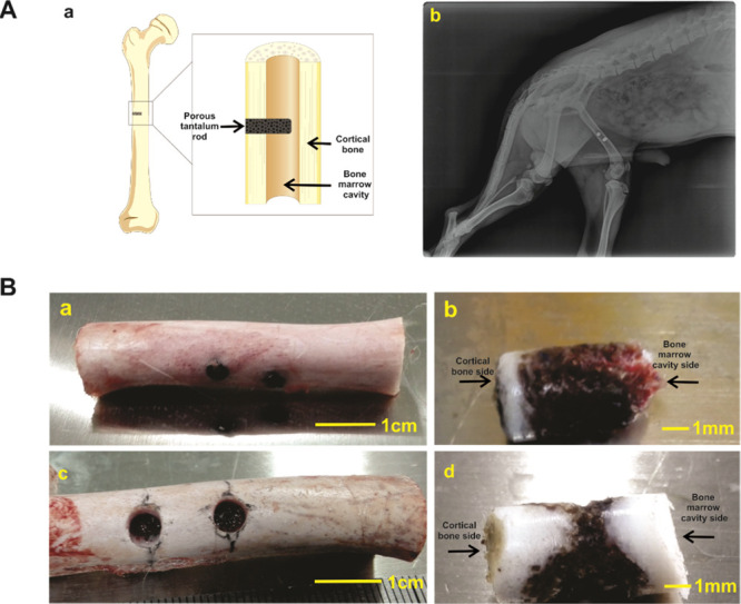
Figure 3.

Construction of canine femoral shaft bone defect models and implantation of porous tantalum rod. (A) Graphical diagram for the construction and implantation of porous tantalum rod at canine’s midpiece of the femur shaft (a). X-ray image of 1 month after the implantation (b). (B) Retrieved femoral shaft samples at 3 month (a, scale bar = 1 cm) and 6 month (c, scale bar = 1 cm), and retrieved tantalum rod-bone samples at 3 month (b, scale bar = 1 mm) and 6 month (d, scale bar = 1 mm).
