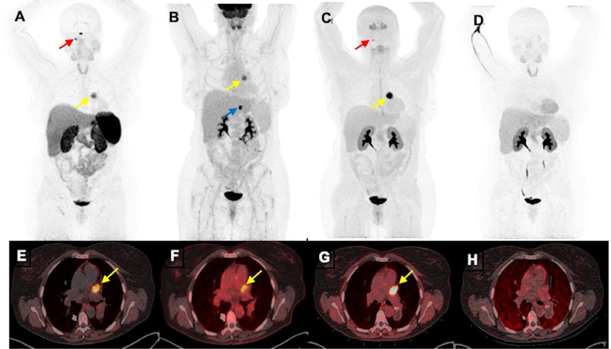
Figure 4.

Functional positron emission tomography/CT (PET/CT) imaging of a cardiac paraganglioma of a woman aged 50 years without mutation in genes encoding subunits of succinate dehydrogenase enzyme. The anterior maximum intensity projection images (A–D) and fused axial PET/CT images (E–H) of 68Ga-DOTA(0)-Tyr(3)-octreotide PET/CT (A, E), 18F-fluorodeoxyglucose (B, F) and 18F-dihydroxyphenylalanine (C, G) PET/CT scans demonstrate a cardiac paraganglioma (yellow arrows) located in between pulmonary trunk and left atrium and superior aspect of left ventricle. However, this lesion lacks avidity on 18F-fluorodopamine (D, H). Furthermore, 68Ga-DOTA(0)-Tyr(3)-octreotide and 18F-dihydroxyphenylalanine PET/CT demonstrate a small right glomus jugulare paraganglioma (red arrows; A, B) and 18F-fluorodeoxyglucose PET/CT alone demonstrates a gastrohepatic nodule (blue arrow, (B)).
