Figure 5.

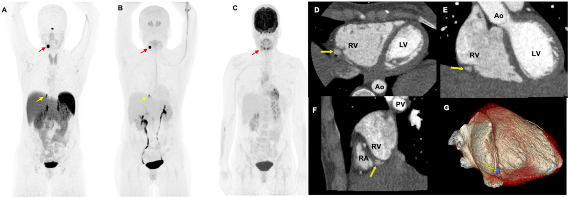
Functional positron emission tomography/CT (PET/CT) and contrast-enhanced cardiac-gated CT imaging of a woman aged 34 years with mutation in succinate dehydrogenase D gene. The anterior maximum intensity projection images (A–C) of 68Ga-DOTA(0)-Tyr(3)-octreotide PET/CT (A) and 18F-dihydroxyphenylalanine demonstrates a pericardiac paraganglioma (yellow arrows; A, B) as well as a left carotid body paraganglioma (red arrows; A, B). However, 18F-fluorodeoxyglucose (C) PET/CT scan demonstrates faint uptake in the left carotid body paraganglioma (red arrow, (C)) but lacks avidity in the pericardiac paraganglioma. This pericardiac paraganglioma was not localised on contrast-enhanced chest CT. The patient further underwent a contrast-enhanced cardiac-gated CT imaging to further characterise this pericardiac mass. The axial (D), coronal (E), sagittal (F) and reformatted three-dimensional images (G) demonstrate a well-circumscribed pericardiac mass (yellow arrows; D–G measuring 0.6×0.7×1.1 cm. This mass is located adjacent to mid-distal right carotid artery (*, (D)) without impingement of vessel lumen. The mass is outside the right ventricular free wall (yellow arrows, (D–F)) in the basal portion of the right atrial-ventricular groove (F) and within the pericardium. Ao, aortic root; LV, left ventricle; PV, pulmonary vein; RA, right atrium; RV, right ventricle; asterisk (*), right carotid artery.
