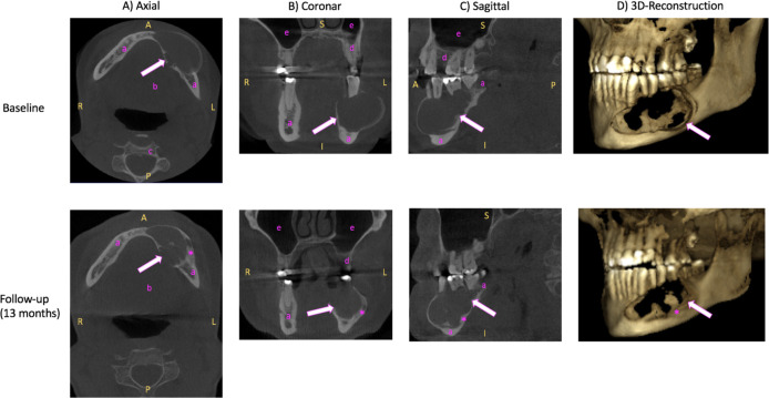
Figure 1.
Three-dimensional (3D) cone-beam computed tomography (CBCT) of the mandible. (A) Axial, (B) coronal, (C) sagittal view and (D) 3D reconstruction of unenhanced CBCT images of the ameloblastoma (arrow) in the left mandible at baseline (2 years before surgery, upper line) and at follow-up 1.5 years after the baseline examination (lower line). Baseline (upper row): Ameloblastoma (arrow) appears as roundish, sharply defined hypodense lump. Follow-up (lower line): Compared with baseline, the ameloblastoma (arrow) appears more sclerosed (asterisk) at the edges with relative size constancy. R, right; L, left; A, anterior; P, posterior; S, superior; I, inferior; a, mandible; b, floor of the mouth; c, vertebral body; d, maxilla; e, sinus maxillaris.

