
Figure 3.
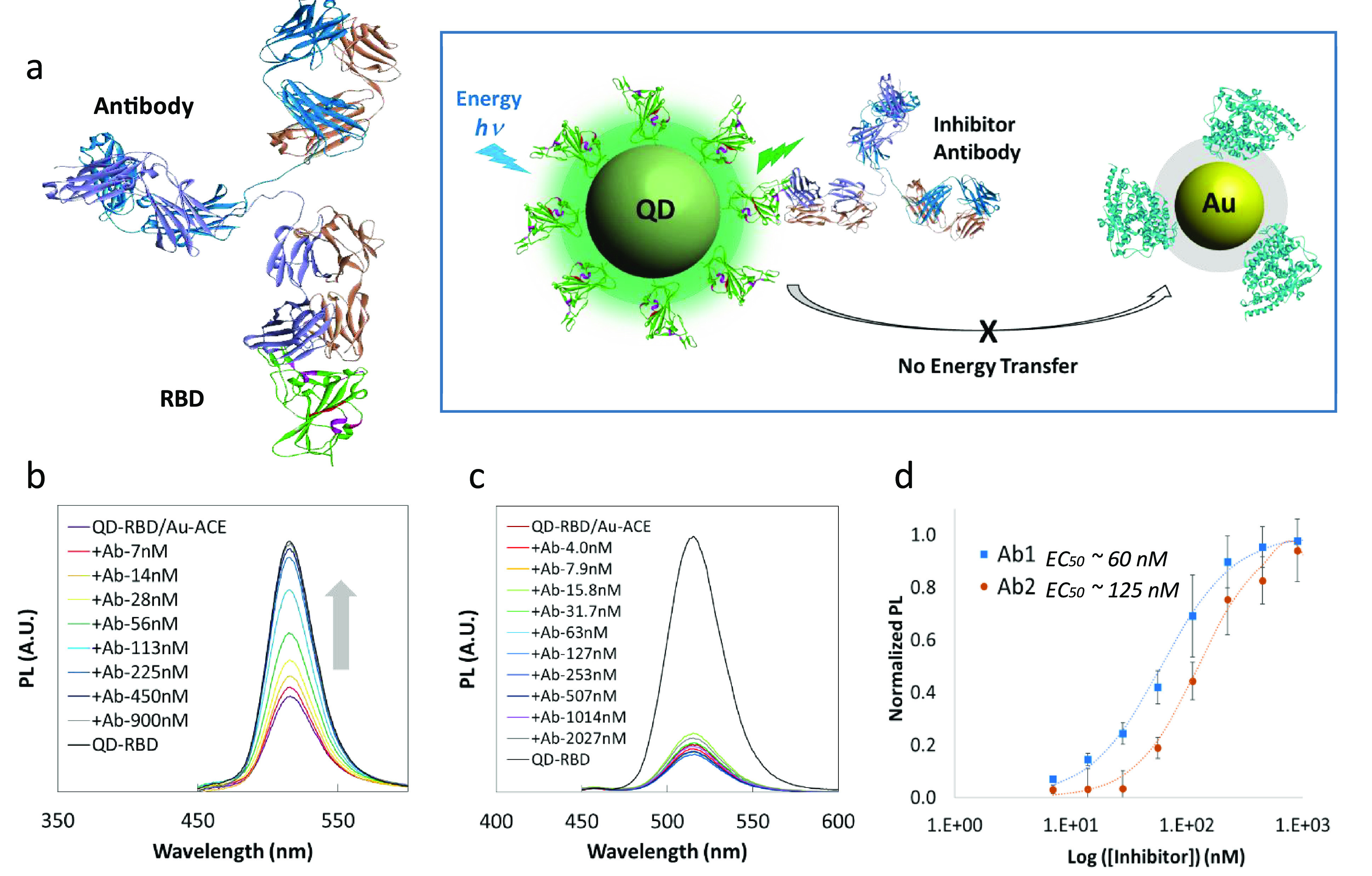
NP-based inhibition assay. (a) Left: The structure of neutralizing antibody (top) bound to SARS-CoV-2 Spike RBD (bottom, green). Right: Schematic diagram of the inhibition assay depicting blocking of the interaction between RBD and ACE2 and the resulting inhibition of energy transfer from QD to AuNP. (b) PL recovery of QD514-RBD in the presence of neutralizing antibody Ab1. (c) Inhibition test using anti-Spike antibody without neutralizing ability, showing almost no PL recovery of QD514-RBD. (d) Calculated EC50s for neutralizing antibodies Ab1 and Ab2 were 60 nM and 125 nM with R2 > 99%, respectively.