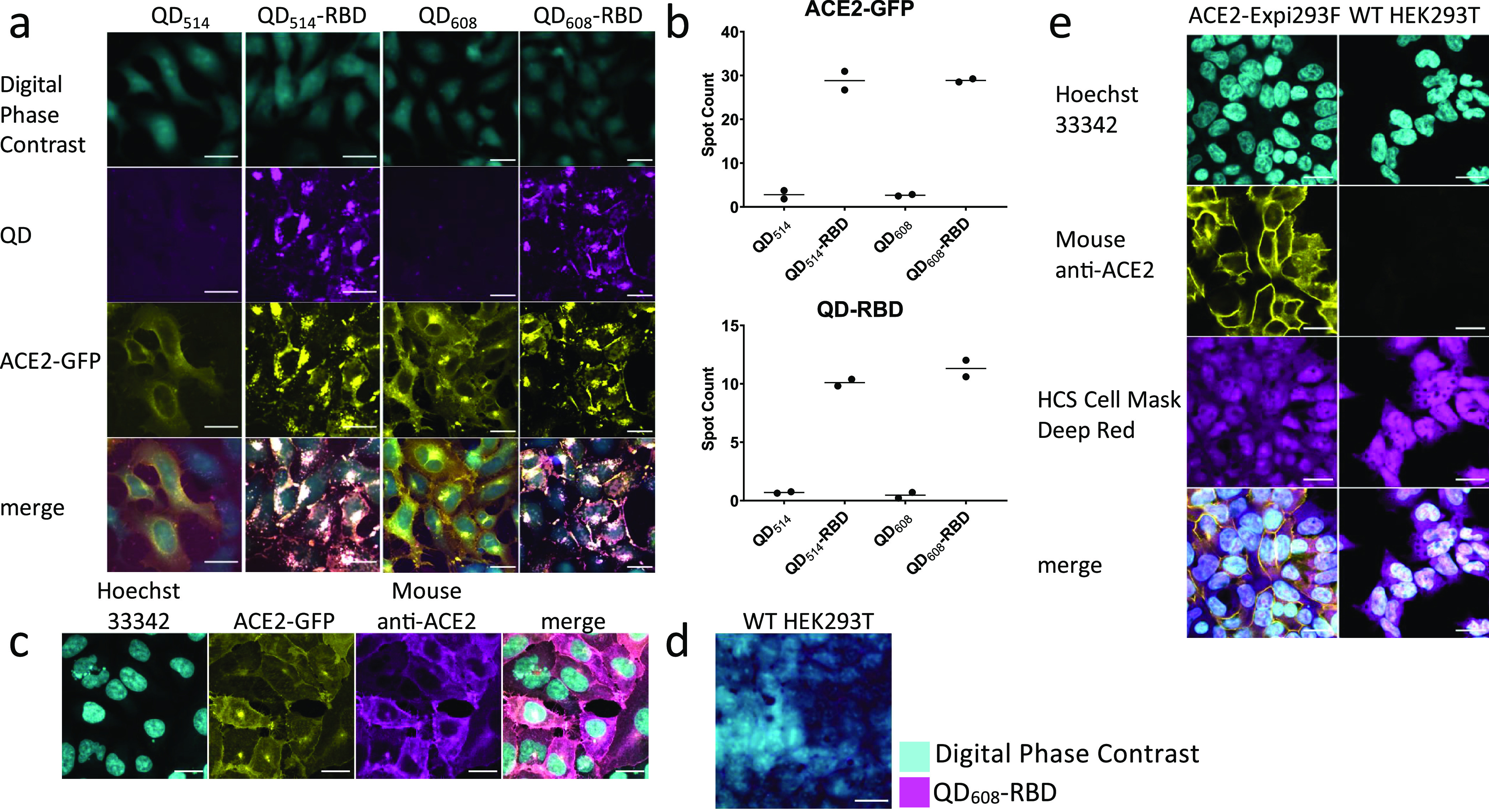
Figure 4.

Quantum dot-conjugated Spike-RBD domain induces translocation of ACE2 and internalizes into cells. (a) Representative image montage of ACE2-GFP (yellow) HEK293T clone 2 treated with 100 nM QD514-RBD (magenta) and QD608-RBD (magenta). Digital phase contrast (cyan) was used during live-cell imaging to identify cell somas. (b) High-content analysis averages of spot count for QD514-RBD and QD608-RBD and ACE2-GFP. N ≥ 400 cells from duplicate wells. (c) Representative image montage of immunofluorescence staining for ACE2 in ACE2-GFP HEK293T cells. Cells were stained with Hoechst 33342 for nuclei (cyan), mouse anti-ACE2 antibody (yellow), and HCS Cell Mask Deep Red for whole cell fill (magenta). N = 9 fields each from 3 triplicate wells. (d) WT HEK293T cells were treated with 100 nM QD608-RBD. Digital phase contrast in cyan and QD608-RBD in magenta. (e) Representative image montage of ACE2-Expi293F and WT-HEK293T cells stained with Hoechst 33342 (cyan), mouse anti-ACE2 antibody (yellow), and HCS Cell Mask Deep Red (magenta). N = 3 triplicate wells. Optimem I alone used as control. Scale bar, 25 μm.
